
Treatment Insights
Submandibular Gland Botox: Anatomy, Injection Location, and Depth
Muscles involved in swallowing and voice production, along with blood vessels, lie close to the submandibular glands. I’ll explain how anatomy and a 2024 ultrasound study help guide injection location and depth.

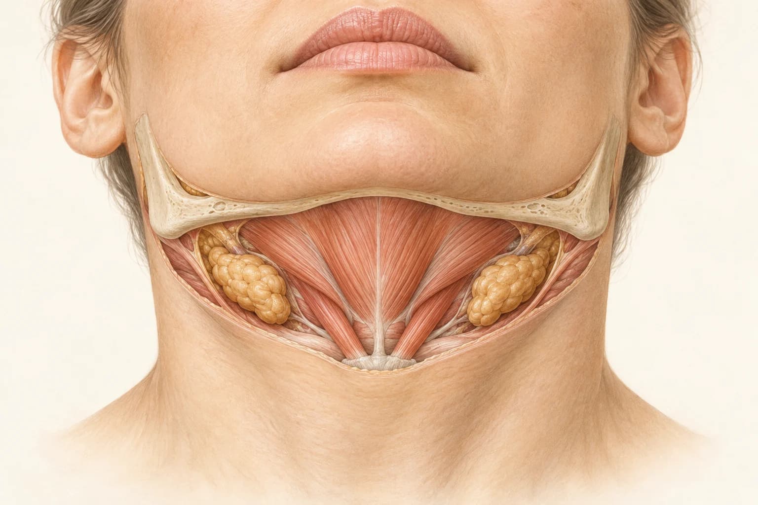
The key point
When planning submandibular gland Botox, I assess the gland’s location, its depth beneath the skin, and the surrounding muscles and blood vessels. I use anatomical landmarks and ultrasound study measurements to plan treatment around each person’s facial structure and body build.
- How the submandibular glands relate to the muscles around the mouth and neck
- The landmarks proposed and measurements reported in an ultrasound study of 42 adults
- My treatment principles: assessing the cause and planning the injection site, depth, and volume
Where are the submandibular glands?
Hello, I am Dr. Sijin Park, the director of Layer Clinic. In the previous article, I discussed the effects of submandibular gland Botox, who it is suitable for, and its possible side effects. Here, I’ll explain the gland’s anatomy and the evidence behind the choice of injection location and depth.
There are three types of major salivary glands: the parotid glands in front of the ears, the submandibular glands beneath the jaw, and the sublingual glands beneath the tongue.
The submandibular gland lies within the submandibular triangle. It has superficial and deep parts on either side of the mylohyoid muscle. The superficial part is the main contributor to a rounded contour beneath the jawline.
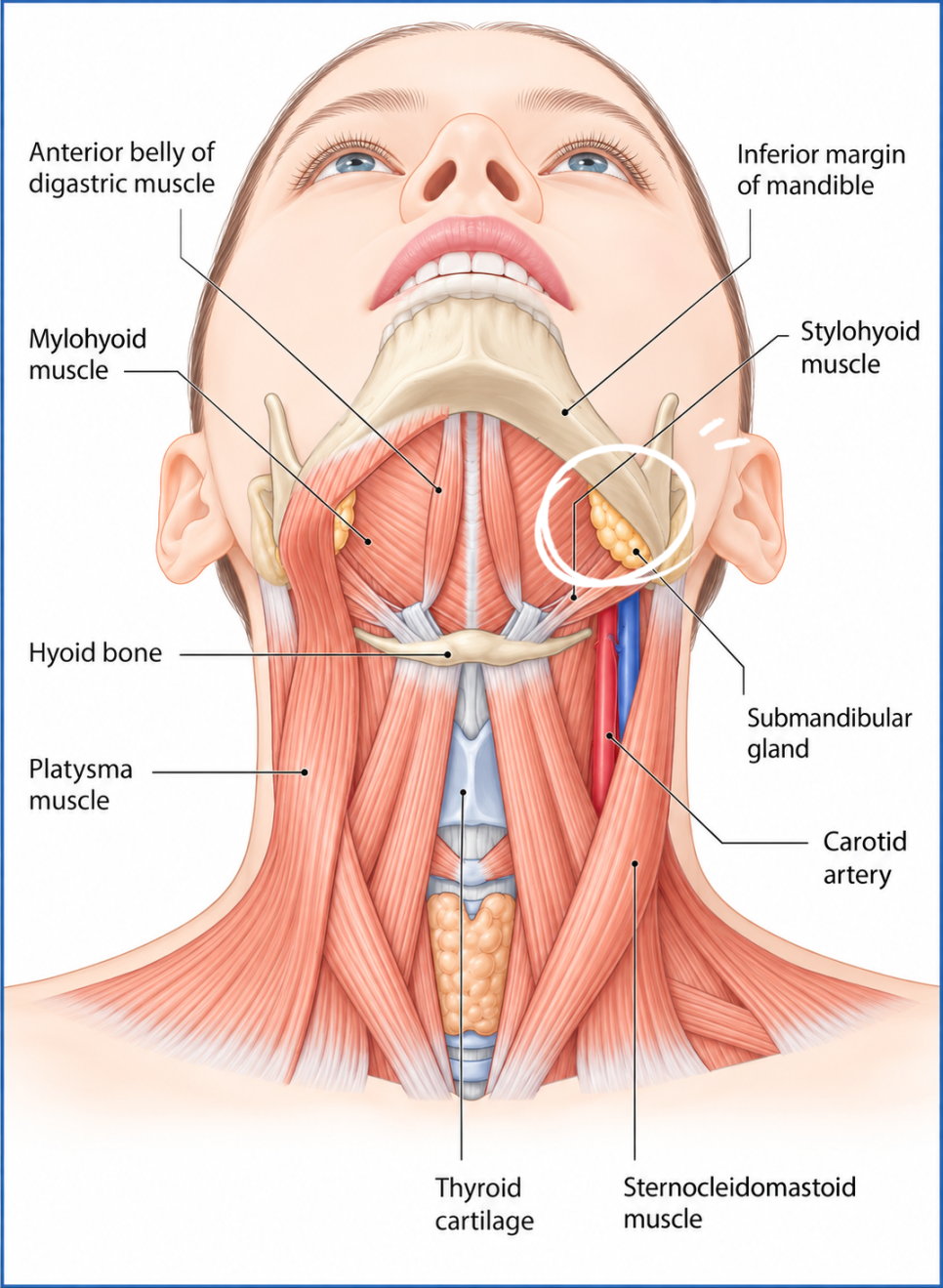
Why the surrounding muscles matter
The submandibular gland does not sit in isolation just beneath the skin. Looking at the layers of tissue from the surface inward helps explain why location and depth matter.
Platysma
The broad platysma lies in the most superficial layer. This superficial neck muscle is supplied by a branch of the facial nerve.
Digastric muscle
This muscle connects the lower jaw and the hyoid bone. It helps lower the jaw to open the mouth or raise the hyoid bone, and it also plays a role in swallowing.
Stylohyoid muscle
This muscle connects the styloid process to the hyoid bone. By raising the hyoid bone, it helps with swallowing and speech.
Mylohyoid muscle
This muscle originates along the mylohyoid line on the inner surface of the lower jaw and attaches to the median raphe and hyoid bone. It forms the floor of the mouth and helps lower the jaw and raise the hyoid bone and tongue.
These nearby muscles are involved in opening the mouth, swallowing, and voice production. If the medication affects surrounding muscles rather than the gland, it can cause discomfort in everyday activities. This makes it important to accurately identify the gland’s location and depth.
What the 2024 ultrasound study examined
Various landmarks have been proposed for locating the submandibular gland. I’ll focus on a study by an anatomy research team at Yonsei University College of Dentistry, published in Dermatologic Surgery in 2024.
Yi and colleagues used ultrasound to examine both submandibular glands in 42 adults and confirmed their location in two cadaveric specimens. The study measured the glands’ depth beneath the skin, thickness, and position relative to blood vessels.
A landmark: the vertical line from the outer corner of the eye
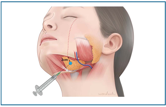
The study used a vertical line extending downward from the outer corner of the eye, or lateral canthus as a landmark. The researchers examined the gland’s position in relation to this line and the lower border of the mandible.
On ultrasound, this line passed near the anterior third of the gland in around 80% of cases. Rather than targeting only the center of the gland, the researchers proposed an injection site using this landmark while taking the position of blood vessels into account.
Depth and thickness measured by ultrasound
The researchers took separate measurements at the gland’s thickest point and where the vertical line from the outer corner of the eye crossed the gland.
| Measurement site | Depth from skin to gland | Gland thickness |
|---|---|---|
| Thickest part of the gland | 4.63 ± 0.76 mm | 11.12 ± 2.46 mm |
| Where the lateral canthus line crosses the gland | 5.53 ± 1.83 mm | 8.73 ± 1.64 mm |
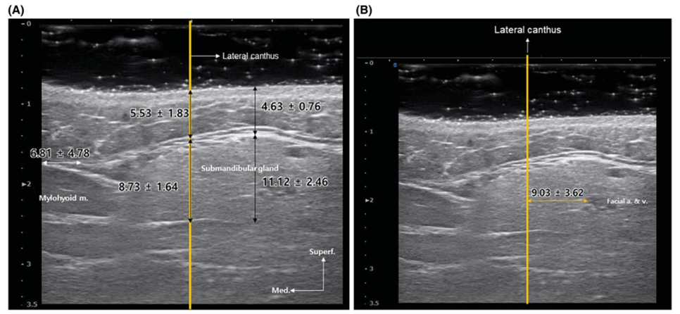
In this study, the gland began approximately 5 mm beneath the skin and was about 9–11 mm thick. The researchers explained that it could be reached with a 0.5-inch needle, approximately 13 mm long. Needle length and actual injection depth should be considered separately.
Depth can also vary with body build and body mass index (BMI). Even when the same landmark is used, depth must be determined according to the gland’s actual position and the person’s facial structure and body build.
Blood vessels just below the jawline
In 37 of the study’s 42 participants, the facial artery and vein ran immediately beneath the mandible. The researchers took these vessels into account when proposing a site approximately 2 cm below the lower mandibular border.
When choosing an injection site, I first distinguish the gland from the blood vessels just below the jawline. I then consider the lateral canthus line, the lower mandibular border, findings on palpation, and depth together to select the site.
Layer Clinic’s approach to submandibular gland Botox
I provide both the consultation and treatment myself. For submandibular gland Botox, I follow these steps.
- Identify the cause: I palpate the area to determine whether fat, muscle, or the gland is creating the fullness.
- Confirm the location: I use the lateral canthus line and jawline as landmarks, then carefully palpate the area to assess the gland’s position relative to blood vessels and select the site.
- Adjust the depth: I adjust the depth to the person’s facial structure and body build.
- Start with a small volume: I inject a small volume at a high concentration to reduce spread to surrounding tissues.
The cause of fullness under the chin differs from person to person. I first determine whether the fullness comes from fat, muscle, or the gland. I then choose the location and depth based on the anatomy, taking blood vessels and the muscles involved in swallowing into account.
Original article and references
- Submandibular Gland Botox: Technique and Anatomical Principles — Layer Clinic blog, October 19, 2026.
- Yi KH, et al. Ultrasonographic Study of the Submandibular Gland for Botulinum Neurotoxin Injection. Dermatologic Surgery. 2024;50(9):834–837. DOI.
What does this mean for your skin?
Tell us about your concerns and treatment history. Your doctor can discuss the options with you.
Ask on WhatsApp